
Figure 1.
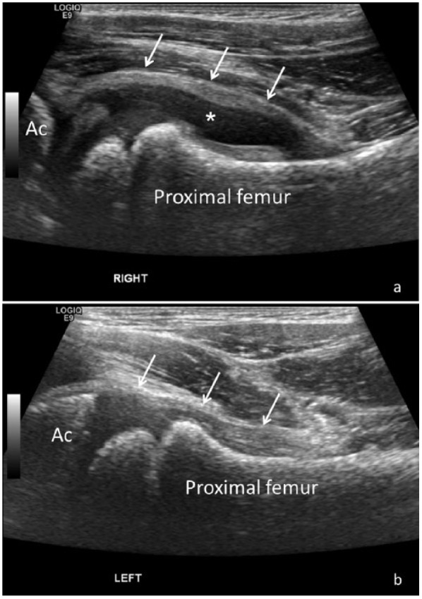
Sonographic evaluation of the hips in a pediatric patient with right hip pain and clinical suspicion for juvenile inflammatory arthritis. (a) Longitudinal ultrasound (US) image of the right hip reveals distension of the joint capsule (white arrows) by a large joint effusion (*), in keeping with synovitis. (b) Comparative longitudinal US of the left hip demonstrates a normal hip joint with a decompressed joint capsule (arrows). Ac, acetabulum.