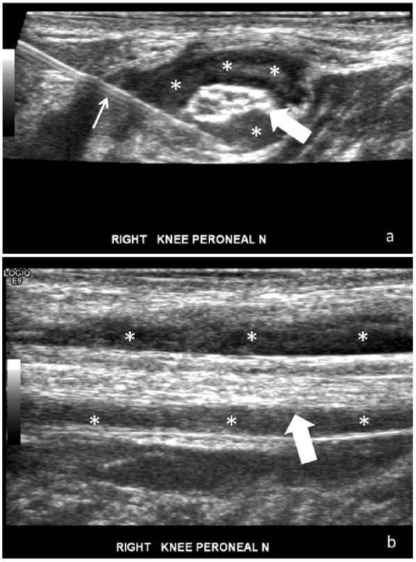
Figure 8.

Ultrasound (US)-guided hydrodissection of the common peroneal nerve in a patient with radicular dorsolateral leg and foot weakness. (a) Transverse US image just proximal to the fibular head shows large volume injectant (*) circumferentially dissecting away tissues around the common peroneal nerve (block arrow) during hydrodissection. (b) Longitudinal US image of the common peroneal nerve (block arrow) after hydrodissection demonstrates injectant (*) along the superficial and deep surface of the nerve. Small arrow, 25-gauge needle.
