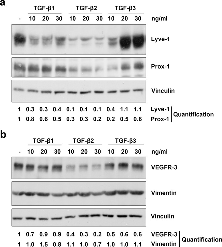
Fig 3. TGF-β1, -β2 and -β3 reduce lymphatic marker expression in LECs.
Primary human LECs were treated with 10, 20 or 30 ng/ml TGF-β1, -β2 and -β3 for 72 hours (a) or 100 hours (b). Untreated cells served as a control. Lysates were prepared and analysed by Western blot using antibodies specific for Lyve-1, Prox-1, VEGFR-3 or vimentin. Vinculin served as loading control. The experiment was performed twice with equivalent results. For densitometry evaluation, protein bands were analysed using the software ImageJ. Bands for the Prox-1, Lyve-1, vimentin and VEGFR-3 proteins were normalized to the corresponding loading control and are displayed as the expression level relative to the untreated control samples.

