
Fig. 7.
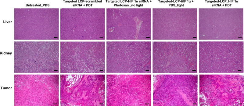
Hematoxylin and eosin-stained tissue sections from SCC4 xenograft tumor-bearing mice after treatment with targeted LCP-HIF1α siRNA. SCC4 xenograft tumors reached 500–800 mm3 and then mice were intravenously injected on days 0, 1 and 2 with PBS or 6 μg LCP carrying either scrambled siRNA or HIF1α siRNA followed by photosan and light treatment on day 3. No major changes were observed in LCP-HIF1α siRNA treated liver and kidney tissue compared to scrambled siRNA or untreated tissue on day 10 after the start of treatment. However, major hollow areas were seen in tumor tissues treated with HIF1α siRNA. Bar standards for 100 μm.