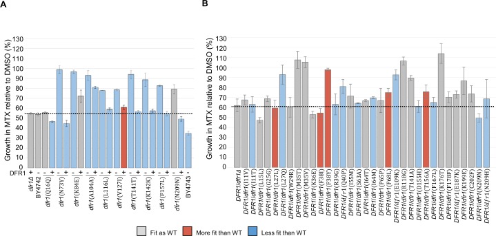
Fig 3. Validation of methotrexate resistant dfr1 mutations.
The average fitness of the candidate dfr1 haploid (A) and diploid (B) mutants upon exposure to MTX (1 mM sublethal dose) or DMSO solvent (2% v/v) were evaluated over 24 hours in a Tecan shaker-reader at 30°C. Mutant alleles expressed in the presence of a wild-type DFR1 copy are listed with (+). Mutants are colored according to their relative doubling time in comparison to the wild-type strains: BY4742 (WT, A) and DFR1/dfr1Δ (WT, B) with: blue for longer doubling time (Slower than WT), grey for comparable doubling time (Fit as WT), and red for shorter doubling time (Faster than WT). The average growth under MTX conditions of the control strains are indicated with a dashed line. Error bars indicate standard error, n = 3.

