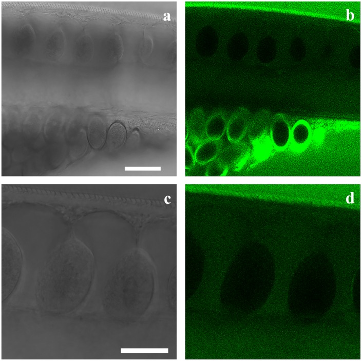
Fig 8. Transmission image (Fig 8a and 8c) and confocal section (Fig 8b and 8d) through an adult Trichuris muris at the junction between the thin anterior and the thick posterior part of the worm after incubation in agarose (1%) with 6-(N-(7-Nitrobenz-2-oxa-1,3-diazol-4-yl)amino)-6-Deoxyglucose (6-NBDG) (200 μM) for approximately 40 minutes.
Note the eggs are located ventrally near the vulva, which is itself located at the border between the thin anterior and the thick posterior part of the worm (Fig 8a and 8b). The bacillary band was not visible in this area and the stichocytes and the connective membranes were devoid of 6-NBDG. Scale bar: 50 μm. High magnification of the stichocytes and the connective membranes devoid of 6-NBDG (Fig 8c and 8d). Scale bar: 30 μm.

