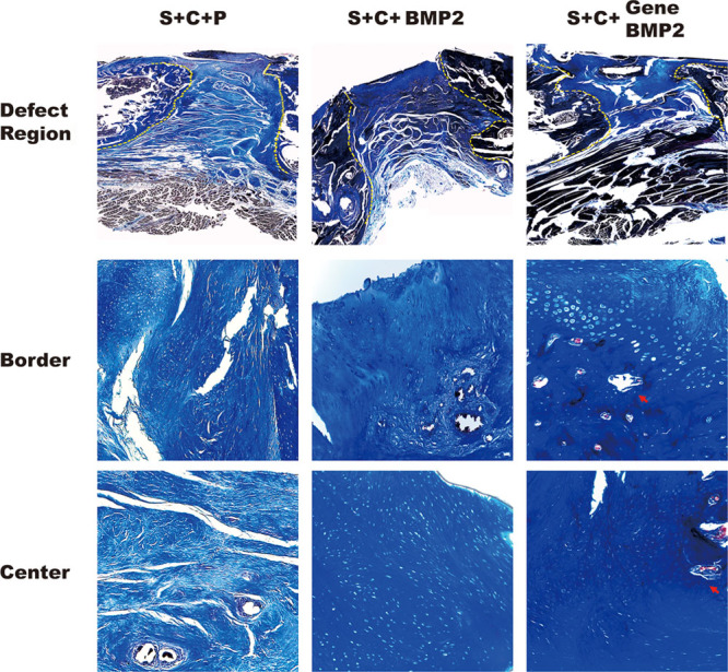
Fig. 2.

Histological analysis of new bone formation. CDM group: BMMSCs were encapsulated in the PEG-PLLA scaffold and cultured in CDM. The border between old and new bone showed chondrocytes and little vessel formation. S + C + BMP2 group: chondrocytes were present at both the border and the center of the bone defect area. S + C + gene BMP2 group: Cartilage formation was present everywhere. The ossification occurred in the border and center of the defect area. New bone was observed both in the center and border of the defect. The bone defect area lies between the yellow dashed lines. The images of border and center region were at larger magnification (magnification, 100×).
