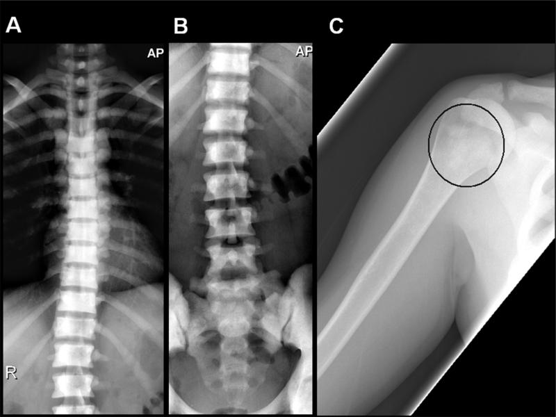
Figure 2.
Anteroposterior radiographs of the thoracic and lumbar spine (A,B) demonstrate severe diffuse osteosclerosis of the vertebral bodies with loss of normal cortical-medullary differentiation. Imaged portions of the ribs and pelvis shows similar findings. Anteroposterior radiograph of the right proximal humerus (C) demonstrates patchy intramedullary sclerosis of the proximal shaft (ellipse), different in character from the diffuse marrow replacement of the axial skeleton.

