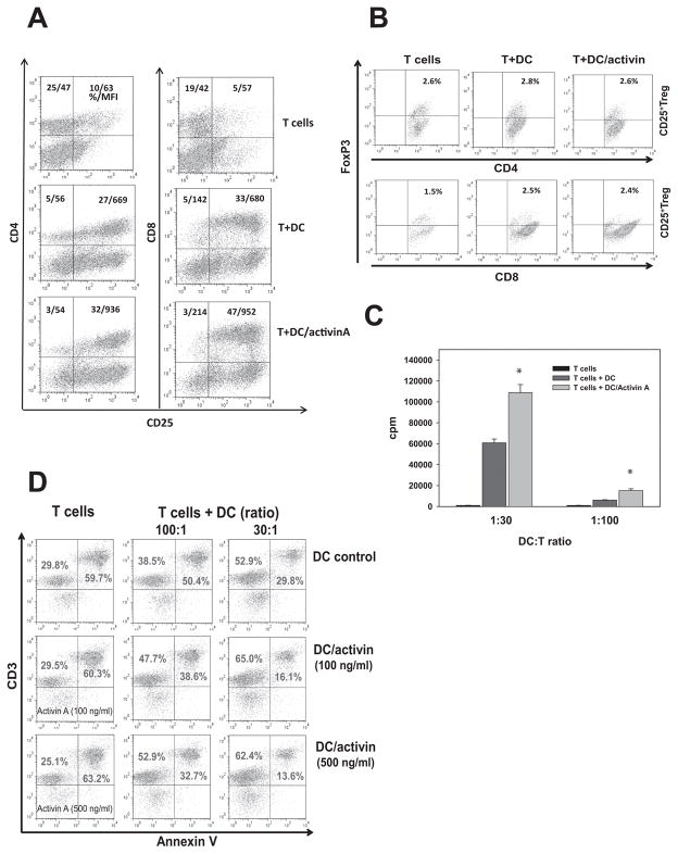
Figure 2. Activin A-treated DC up-regulate activation (A), proliferation (C) and survival (D) of T cells in vitro.
Bone marrow-derived Day5 DC were treated with ActA (100 ng/ml, 48h) and mixed with ConA (2.5μg/ml) pre-activated splenic T-cells for 48h. Expression of CD25 (A) on CD4+ and CD8+ T-cells and expression of FoxP3 (B) on gated CD25+CD4+ and CD25+CD8+ T-cells was determined by flow cytometry. The results of one representative out of three independent experiments are shown. C. Proliferation of T-cells mixed with control and ActA-treated DC at different ratios was assessed by 3H-thymidine incorporation and expressed as a count per minute (cpm). *, p<0.05 (ANOVA, mean ± SEM, n=3). D. Control and ActA-treated (100 and 500ng/ml, 48h) DC were co-incubated with ConA pre-activated and washed T-cells at different ratios. Untreated and ActA-treated T-cells without DC (left column) served as a control. The level of CD3+ T-cell apoptosis was measured by Annexin V binding. The results from a representative experiment are shown (n=3).

