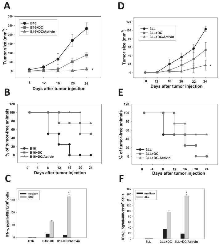
Figure 5. Activin A augments the antitumor efficacy of DC in melanoma and lung carcinoma models.
B16 (A,B,C) and 3LL (D,E,F) tumor cells (0.05×10⁶) were inoculated s.c. in flank of C57BL/6 mice in three groups (5–7 mice/group) on Day1: (i) control (tumor cells only), (ii) control DC, (iii) ActA-DC. Control or ActA-treated DC (100ng/ml, 48h) (1×10⁶cells/mouse) were injected s.c. on Day1 and Day7. The tumor size was expressed as the tumor area (mm2) and shown as mean±SEM (A, D). *, p< 0.05 vs groups (i) and (ii) (Two way ANOVA, n=3). Survival of animals was also determined (B,E). For evaluation of tumor-specific CTL, splenic T-cells were isolated on Day24 and stimulated with medium (control) or irradiated (30,000Rad) B16 or 3LL cells at 5:1 T-cell:tumor cell ratio. Supernatants were collected 48h later, and IFN-γ levels were determined by ELISA. Supernatants from non-stimulated T-cells were used for assessing spontaneous cytokine production (C,F) (*, p<0.05 vs. control DC, Two way ANOVA, n=3).

