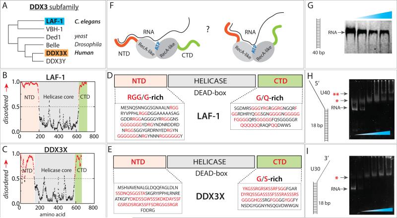
FIGURE 1. Domain composition of LAF-1 and DDX3X.
(A) DDX3 subfamily phylogeny. (B, C) Analysis of intrinsically disordered domain of LAF-1 and DDX3X. (D, E) Composition both proteins containing intrinsically disordered N- and C-terminal domains and central helicase. (F) Unknown protein-RNA interface between LAF-1 and RNA. (G) EMSA showing no LAF-1 binding to 40bp dsRNA. (H, I) EMSA demonstrating LAF-1 binding to partially duplexed, partially single strand RNA with 5’ tail (H) and 3’ tail (I). Single and double red star denote one and two unites of LAF-1 bound band, respectively. See also Figure S1.

