
Fig. 1.
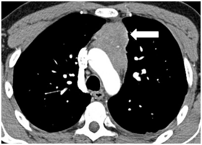
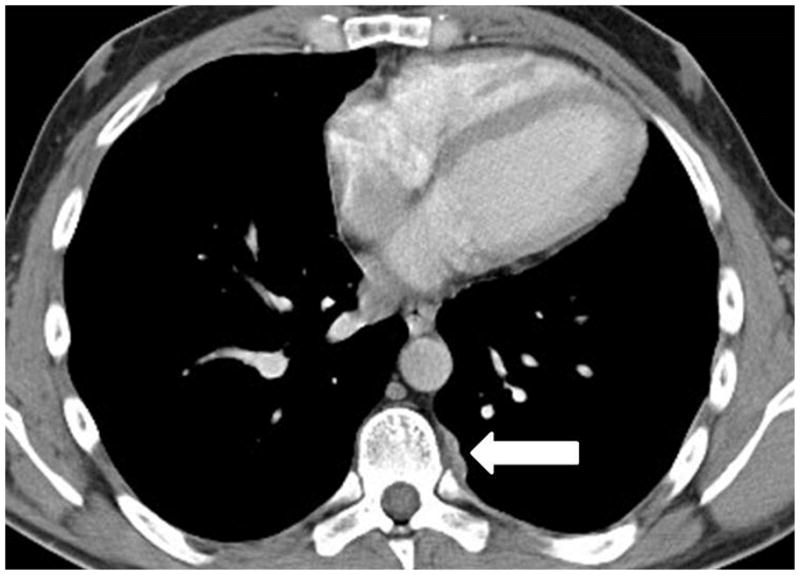
A 35-year-old man with thymoma type B3. (a) Preoperative chest CT demonstrated a large anterior mediastinal mass with a lobulate contour, abutting the aortic arch and infiltrating the surrounding fat. The patient was treated with neoadjuvant chemotherapy and radical thymectomy. (b) Follow-up CT 22 months after surgery demonstrated a new pleural lesion on the left (arrow), which was surgically resected and histopathologically confirmed to be metastatic thymoma.