
Fig. 2.
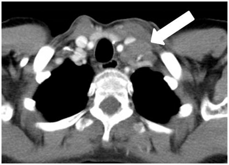
A 44 year-old woman with myasthenia gravis and thymoma type B3, which was surgically resected. Follow-up chest CT after 69 months of surgery demonstrated an enlarged left supraclavicular lymph node, which was histologically confirmed as metastatic thymoma.