
Fig. 4.
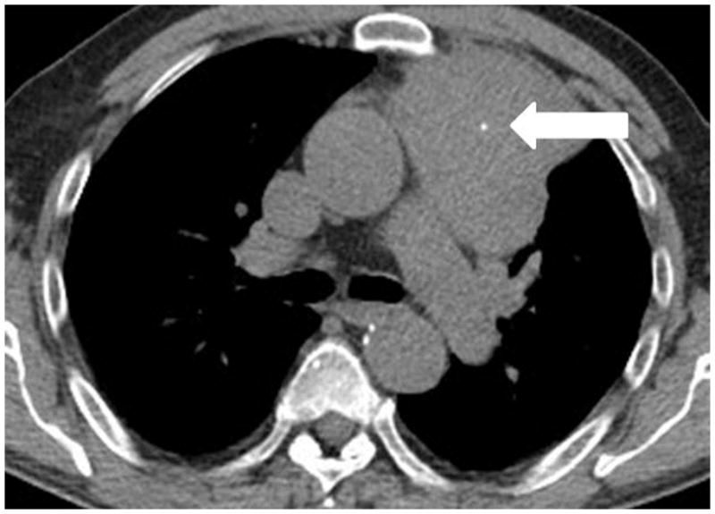
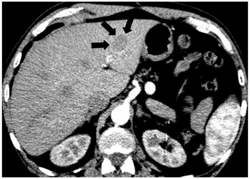
A 64-year-old man with thymic carcinoma. (a) Preoperative CT demonstrated a large anterior mediastinal mass with a lobulate contour abutting the pleura, with a focus of calcification (arrow). The patient underwent radical thymectomy followed by chest radiation with concurrent chemotherapy with two cycles of etoposide and cisplatin. (a) Follow-up chest CT 25 months after surgery demonstrated a new liver lesion in the left lobe (arrows). The liver biopsy was performed and metastasis from thymic carcinoma was confirmed.