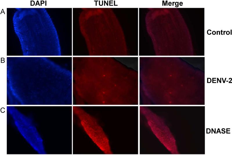
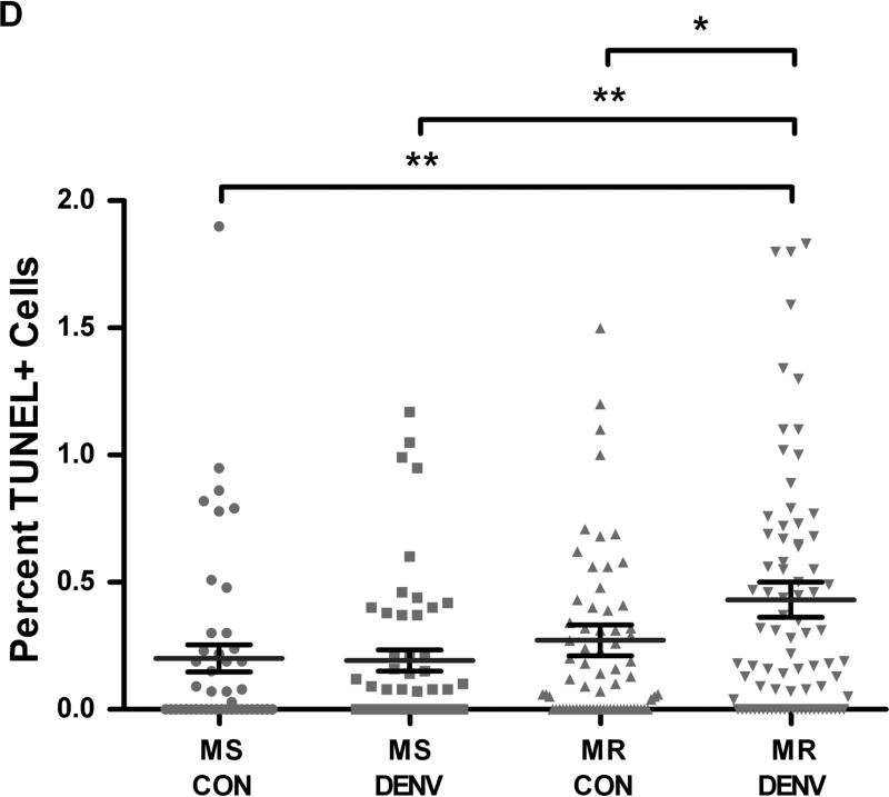
Fig 2. DENV-2-infected midguts from refractory strain (MR) of Ae. aegypti show increased apoptotic cells.
MR and MS females 4-5 days post eclosion were fed either (A) control or (B) DENV-2 JAM1409-supplemented artificial blood meals. At 48 h PBM midguts were dissected out and nuclei were stained with DAPI (blue) and apoptotic cells were stained by TUNEL (red). (C) As a positive control, midguts were incubated with DNAse for 30 min and then stained for TUNEL. Depicted images are of MR mosquitoes. Magnification at 40X. (D) Apoptotic nuclei were quantified as the proportion of the total number of cells in each midgut section (n=3 replicates; 16-28 sections per independent replicate). Statistical analysis of percent midgut cells staining TUNEL+ between groups was performed using Mann-Whitney U test. *p<0.05; **p<0.01.


