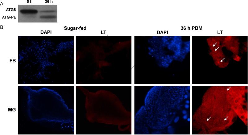
Fig 5. ATG8-PE conjugation and Lysotraker Red staining indicate autophagosome biogenesis following a blood meal.
(A) MR females 4-5 days post eclosion were fed a naïve artificial blood meal. At 0 and 36 h PBM, pools of 5 whole females were tested for presence of the autophagy marker ATG8. ATG8 is cytoplasmic, but becomes conjugated to phosphatidyl-ethanolamine (PE) on the autophagosomal membrane during autophagy. (B) Female fat body and midgut tissues from mosquitoes fed only on sugar water and from mosquitoes 36 h PBM were stained with DAPI and Lysotracker Red. White arrows point to puncta representative of autophagosomes. Images are at 100X magnification.

