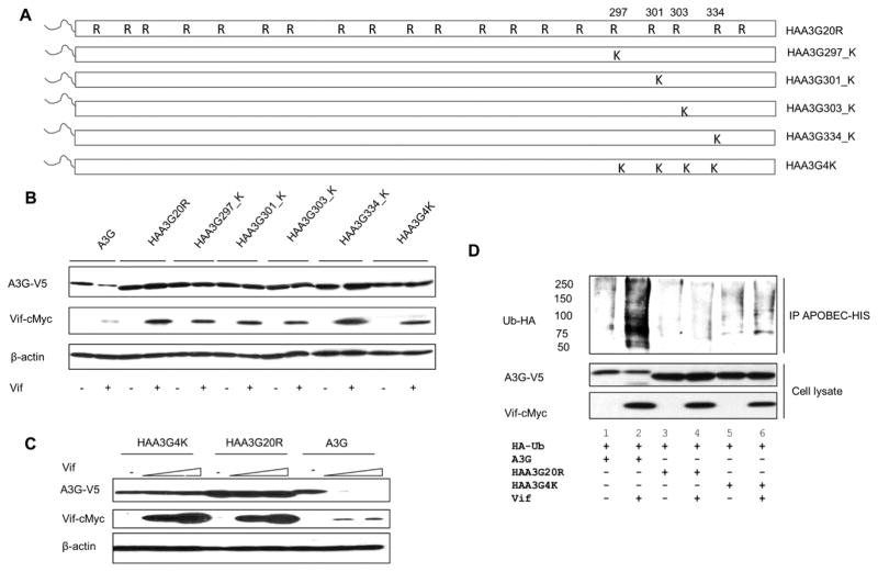
Figure 2. A3G lysine residues 297, 301, 303 and 334 remain Vif insensitive.
(A) Schematic of arginine to lysine reversions generated from A3G lysine-free mutant, HAA3G20R. (B) A3G, HAA3G20R, HAA3G297_K, HAA3G301_K, HAA3G303_K, HAA3G334_K and HAA3G4K were cotransfected with Vif or VR1012 into 293T cells. Forty-eight hours post-transfection, the cells were harvested for Western blot analysis. (C) HAA3G4K, HAA3G20R and A3G were cotransfected with increasing concentrations of Vif (0, 1, 1.5 μg) into 293T cells. At 48 h post-transfection, cells were harvested for Western blotting. (D) 293T cells were cotransfected with A3G, HAA3G20R or HAA3G4K with HA-Ubiquitin-WT and either VR1012 or Vif-c-Myc. At 24 h post-transfection, cells were treated overnight with 10 μM MG132. Ni-NTA beads were used to precipitate A3G under denaturing conditions and analyzed by Western blotting.

