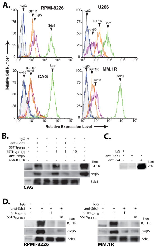
FIGURE 1. Myeloma cells express IGF1R coupled to Sdc1 and αvβ5 integrin.
A. RPMI-8226, U266, CAG and MM.1R human myeloma cells are analyzed by flow cytometry for relative cell surface expression of Sdc1 (B-A38), αvβ5 integrin (P1F6), αvβ3 integrin (LM609) and IGF1R (JBW902). B. Lysates of CAG cells grown in suspension culture are subjected to immunoprecipitation using isotype-matched control mIgG1, B-A38 (Sdc1), P1F6 (αvβ5 integrin) and/or JBW902 (IGF1R) in the presence of SSTNIGF1R (1 μM) or SSTNIGF1R-T (1, 3 or 10 μM). Immunoprecipitates are probed for presence of Sdc1 (B-A38), IGF1R (33255) and αvβ5 integrin (D24A5). C. CAG cell lysates are subjected to immunoprecipitation as in (B) using B-A38 or 44H6 specific for VLA-4 (α4 integrin), and analyzed for the presence of VLA-4. D. Lysates of RPMI-8226 or MM.1R cells are subjected to immunoprecipitation with B-A38 containing no competitor, or 1 μM SSTNIGF1R or 10 μM SSTNIGF1R-T and analyzed as in (B).

