
FIGURE 2. IGF1R activation in myeloma is inhibited by SSTNIGF1R.
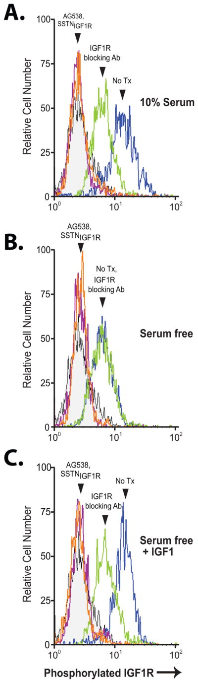
CAG cells cultured in serum-containing medium (A) or serum-free medium for 6 hr (B, C) were treated with 30 μM SSTNIGF1R, 10 μM tyrphostin AG538, or 1.5 μg/mL IGF1R blocking antibody (24–57) versus isotype-matched control mIgG1 for 1 hr in the presence (C) or absence (A, B) of supplementary 100 ng/mL IGF1. Cells were then fixed, blocked, permeabilized, stained with mAb K74-218 specific for pY1131 in the activation loop of IGF1R and analyzed by flow cytometry for levels of activated IGF1R.