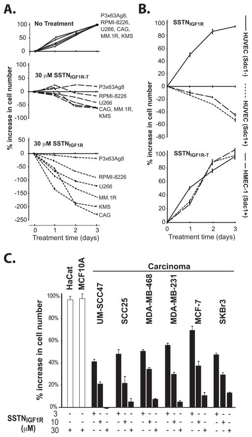
FIGURE 3. SSTN inhibits growth of myeloma, carcinoma and activated endothelial cells.
A. Myeloma cell growth in complete culture medium containing 30 μM SSTNIGF1R or SSTNIGF1R-T is quantified over 3 days and graphed as % increase in cell number relative to untreated cells (set to 100%, top panel); B. Endothelial cells positive (HUVEC (Sdc1+), HMEC-1) and negative (HUVEC(Sdc1−)) for Sdc1 expression were grown in the presence of 30 μM SSTNIGF1R or SSTNIGF1R-T for 3 days and graphed as % increase in cell number compared to untreated cells as in (A); C. Non-transformed HaCaT keratinocytes and MCF10A breast epithelial cells, UM-SCC47 and SCC25 head and neck squamous cell carcinoma, and MDA-MB-468, MDA-MB-231, MCF-7 and SKBr3 breast carcinoma cells grown in 3, 10 or 30 μM SSTNIGF1R for 3 days are graphed as % increase in cell number compared to untreated cells as in (A).

