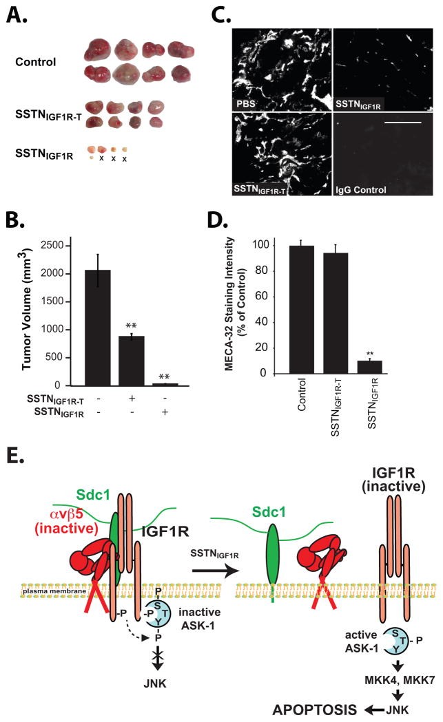
FIGURE 7. SSTN blocks the growth of myeloma xenografts by targeting the tumor cell and angiogenesis.
CAG myeloma cells implanted subcutaneously in immunodeficient nude (nu/nu) mice and are allowed to form palpable tumors for 7–10 days. Mice are then implanted with Alzet pumps delivering 0.25 μL/hr of 4 mM SSTNIGF1R, SSTNIGF1R-T or saline alone for an additional 4 weeks. A. Tumors harvested at the end of the 4-week treatment period (8 per cohort shown; ‘X’ denotes no tumor found). B. Quantification of tumor volume. Treated and untreated cohorts were compared using unpaired one-tail t-test. P-values of less than 0.05 were considered significant (**). Data represent mean plus or minus SEM. C. Representative tumors sectioned and stained with MECA-32 antibody specific for the pan-endothelial cell marker PV1/PLVAP. D. Quantification of overall MECA-32 staining intensity. E. Model depicts capture of inactive αvβ5 integrin and IGF1R via a docking site in the extracellular domain of Sdc1 (21,23), which causes autophosphorylation of IGF1R and binding of inactive ASK1 (with inactivating phosphorylation on Ser83, Ser966 (S) and tyrosines (Y)). Displacement of the integrin and IGF1R from Sdc1 inactivates IGF1R, allow ASK1 activation via autophosphorylation of Thr838 (T), and downstream activation of JNK via MKK4 or MKK7, leading to caspase activation and apoptosis.

