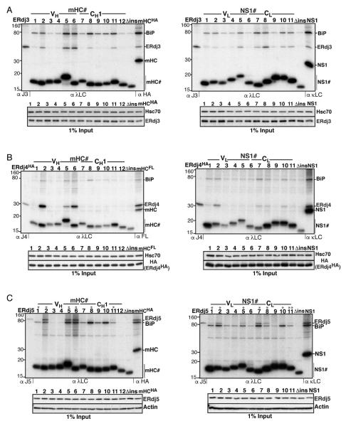
Figure 2. ERdj3 binds frequently throughout mHC and NS1, while ERdj4 and ERdj5 possess fewer, mostly shared binding motifs.
(A) COS-1 cells were co-transfected with ERdj3 and BiP together with the indicated peptide constructs and metabolically labeled, DSP-crosslinked and lysed in RIPA buffer. Peptide constructs were immunoprecipitated with anti-λ antiserum and analyzed by reducing SDS-PAGE. Cells transfected with ERdj3 and BiP alone were immunoprecipitated with antiserum specific for ERdj3 as molecular weight marker, and full-length clients were included as a reference for chaperone binding. Levels of ERdj3 expression in each sample by immunoblotting are shown under the panel. (B) Binding of mHC and NS1 peptides to ERdj4 was performed as in (A), except that HA-tagged ERdj4 was co-transfected and cells were lysed directly in NP40-lysis buffer. (C) Binding of ERdj5 to mHC and NS1 peptides was performed as in (B), except that ERdj5 was co-transfected and the lysis/washing buffers contained 20 mM NEM.

