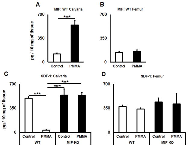
Figure 4.
A PMMA-induced osteolytic lesion has elevated level of MIF and diminished amount of SDF-1 chemokines in vivo. A) and B) Concentrations of MIF detected in calvaria and femur tissues of control (no particles placed) and PMMA particle-induced osteolytic calvarial lesions in WT animals; C) and D) The amounts of SDF-1 in calvaria and femur tissues of control (no particles placed) and PMMA particle-induced osteolytic calvarial lesions in WT and MIF-KO animals. PMMA particles suspended in PBS or PBS alone were placed over calvarial tissues of WT or MIF-KO animals (n=5 animals/group). At day 3, the tissues were dissected and homogenized in PBS buffer containing Tween 20. The supernatants were subjected to a sandwich ELISA. **p<0.01, ***p<0.001.

