Figure 8.
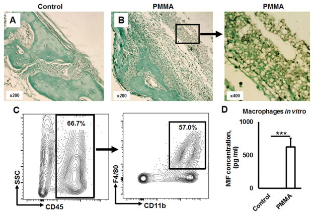
Macrophages of a synovial-like periprosthetic (S-LPP) membrane are the main source of MIF in PMMA particle-induced ostelysis lesions. A) and B) Immunohistochemical detection of MIF in control (no particle placement) and PMMA-particle induced calvaria tissues, respectively. Immuno-histological evaluation of MIF-expression patterns in calvarial tissue was carried out in the WT mice that received the injection of control PBS alone or PMMA suspension in PBS for 7 days prior to the sacrifice. C) The percentage of macrophages gated as CD45+CD11b+F4/80+ cells in dissected peri-prosthetic membranes from the PMMA-induced osteolysis lesion of WT animals (n=5/group) evaluated by multicolor flow cytometry. D) MIF release from bone marrow derived macrophages in response to PMMA particles stimulation in vitro. Bone marrow derived macrophages were stimulated in vitro by PMMA (2.5 × 10⁻³ %, w/v) in the presence of M-CSF (30 ng/ml) for 24 h. The supernatant was collected and the concentration of MIF was evaluated by ELISA. ***p<0.001.

