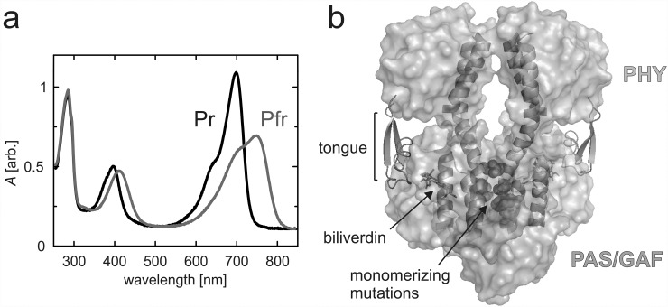
FIG. 1.
UV-Vis spectra and overview structure of the D. radiodurans fragments. (a) UV-Vis spectra of the PAS-GAF-PHYmon in Pr and Pfr states. (b) Domain structure of the photosensory module fragment (PDB code 4Q0J38). PAS and GAF domains are shown in grey and the PHY domain in cyan. The tongue (green) is presented as cartoons, as are the scaffolding helices that form the dimerization interface. The biliverdin chromophore (orange) is presented as sticks, and the monomerizing mutations of three residues which inhibit interactions between the monomers are presented as red spheres.

