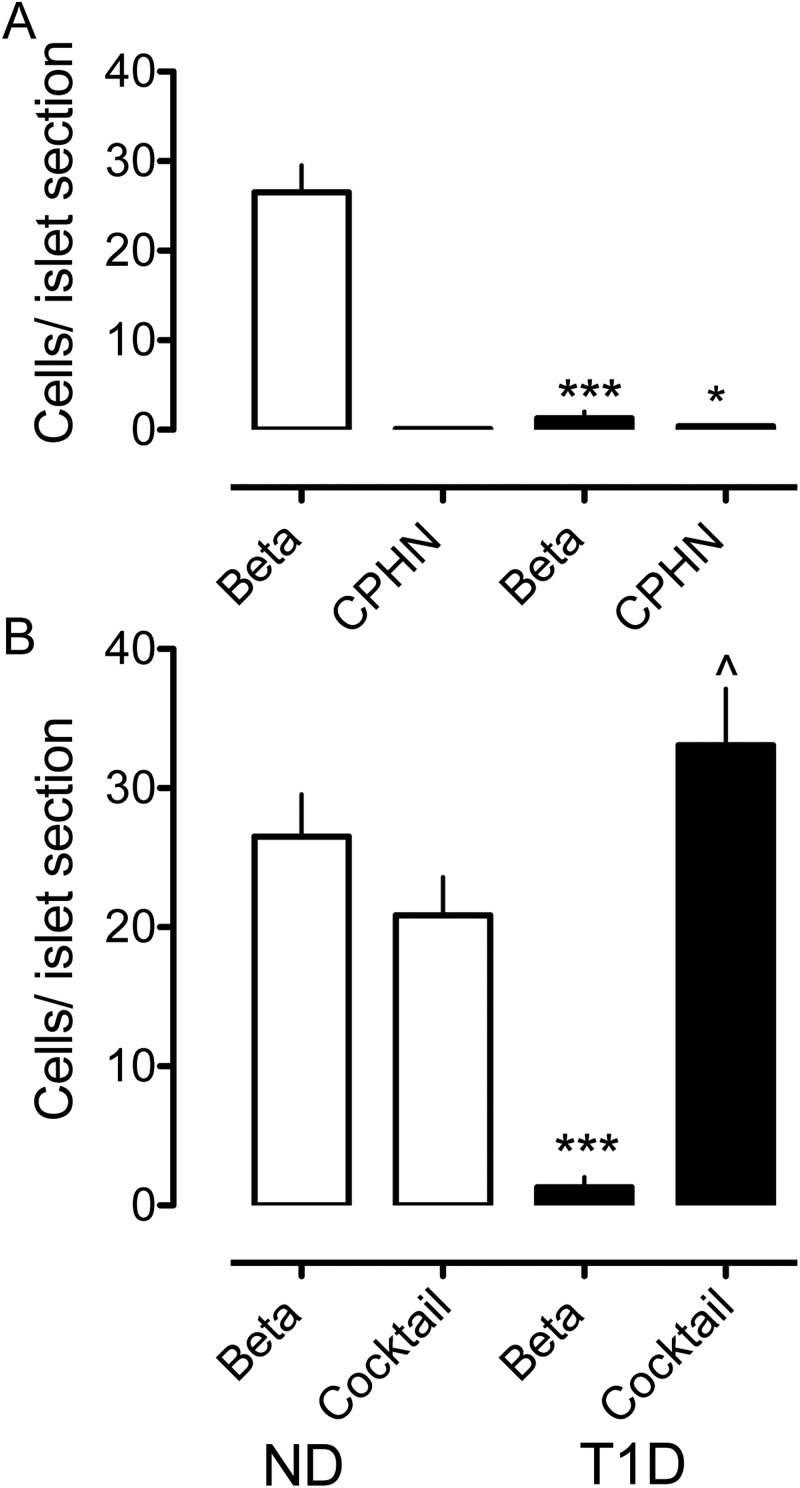
Figure 3.
The impact of nonhormone-expressing endocrine cells on the deficit in β-cell mass in T1D is minimal. The number of cells per islet section of the CPHN cell fraction is very small when compared with the number of the β-cells per islet section in the ND, and the deficit in the β-cells in the T1D donors cannot be accounted for by the increase in the CPHN cells in T1D. A, There is an approximately 95% deficit in the β-cells in the islets from the subjects with T1D (1.31 ± 0.72 vs 26.51 ± 3.03 β-cells/islet, T1D vs ND, ***, P < .0001). The number of the CPHN cells per islet section is increased in T1D (0.40 ± 0.09 vs 0.11 ± 0.05 CPHN cells/islet section, T1D vs ND, , P < .005), but this increase does not account for the β-cell deficit seen in T1D. B, Islet endocrine cells expressing islet hormones other than insulin are increased in T1D (33.10 ± 4.06 vs 20.87 ± 2.74 endocrine cocktail cells/islet section, T1D vs ND, , P < .05). However, the β-cell deficit in T1D (1.31 ± 0.72 vs 26.51 ± 3.03 β-cells/islet section, T1D vs ND, ***, P < .0001) cannot be explained by the conversion of β-cells to other endocrine cells. White bars, ND donors; black bars, T1D donors.

