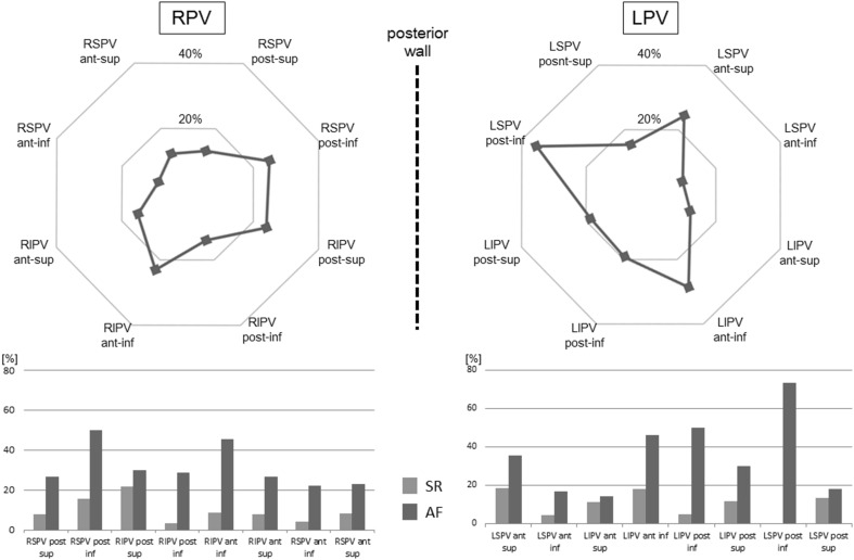
Fig. 7.
The percentage of points with constant contact according to anatomical location. Upper part the proportion of constant contact in all data points. Small percentage of the achievement of constant contact was shown in the ridge area (9.8 % at anterior-inferior of LSPV and 12.2 % at anterior-inferior of LIPV). Lower part the percentage of points with constant contact per each anatomical location according to atrial rhythm. Except for location at the posterior superior of LSPV (p = 0.18), the percentage of constant contact was significantly higher during AF than it was during SR (p < 0.05). The abbreviations used here are same as those used in Fig. 6

