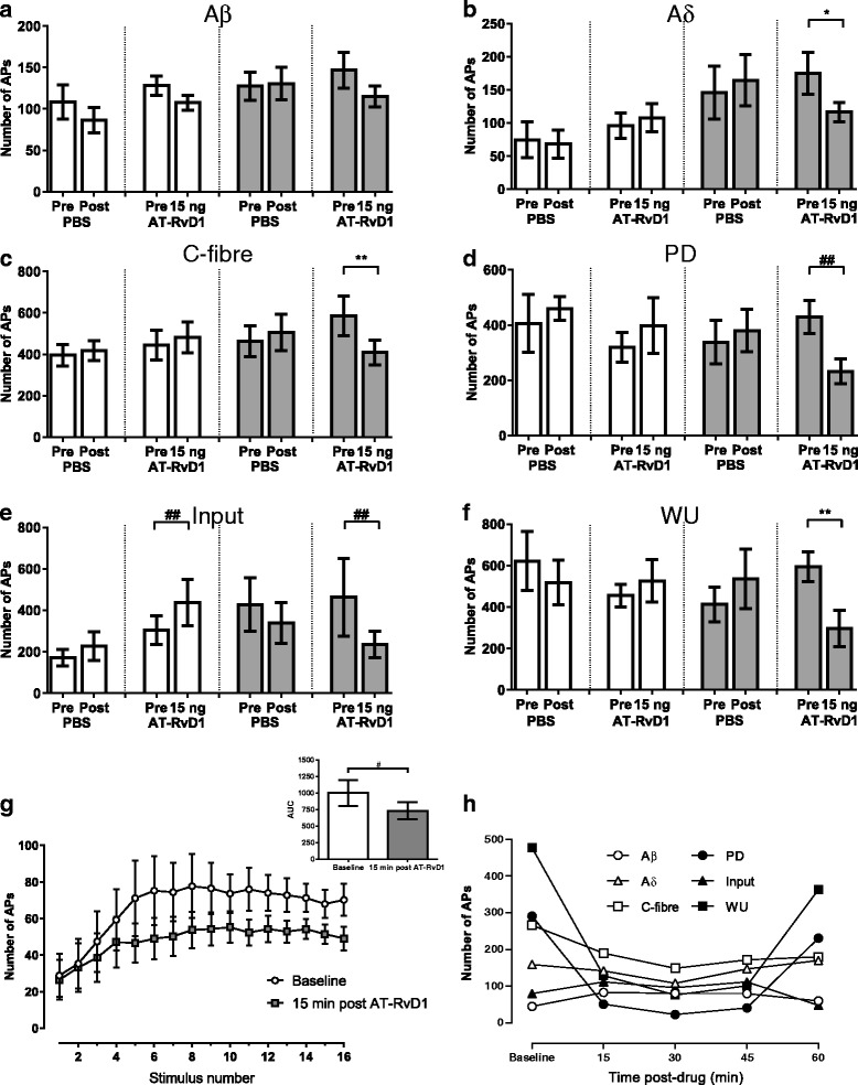
Fig. 2.

Effect of spinal administration of AT-RvD1 on evoked responses of WDR neurons. Spinal administration of AT-RvD1 selectively inhibited nociceptive fibre-evoked responses of spinal WDR neurones in carrageenan-treated rats, which were mediated mainly by FPR2/ALX receptor activation. In saline-treated rats (white bars), electrically evoked responses were not altered by PBS (n = 5) vehicle or AT-RvD1 (n = 10), except the input response which was slightly facilitated by AT-RvD1 (e third and fourth bars). AT-RvD1 (15 ng/50 μl) resulted in a non-significant decrease in Aβ-fibre-evoked responses (p = 0.0781) in carrageenan-treated rats (grey bars, n = 9). Spinal AT-RvD1 significantly suppressed WDR neurone responses evoked by electrical stimulation of Aδ and C fibres (b, c) as well as PD, input and WU (d–f) responses when compared to pre-drug responses. g Illustrates the effect of AT-RvD1 on WU of WDR neurones following 16 electrical stimulations in carrageenan-treated rats. At 15 min post AT-RvD1, the AUC was significantly lowered when compared to baseline (g inset). h Is an example of the electrically evoked responses of a WDR neurone recorded for 60 min following AT-RvD1 application in a carrageenan-treated rat. Data in a–f are mean maximal number of action potentials post drug application. *p < 0.05, **p < 0.01 paired t test. ## p < 0.001 Wilcoxon test, n = 8–10 group, except for saline-PBS n = 5. APs action potentials
