
Fig. 2.
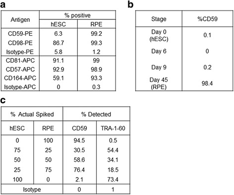
Identification and validation of CD59 expression on RPE cells. a % positive expression of indicated antigens in hESCs and RPE cells as determined by flow cytometry. PE-conjugated or APC-conjugated antibodies together with the respective isotypes were used. b % positive CD59 expression at different stages of the RPE differentiation protocol. c Results of a spiking experiment where different ratios of hESCs and RPE were mixed together and expression of CD59 and the stem cell marker TRA-1-60 was determined by flow cytometry. hESC human embryonic stem cell, RPE retinal pigment epithelium