
Fig. 2.
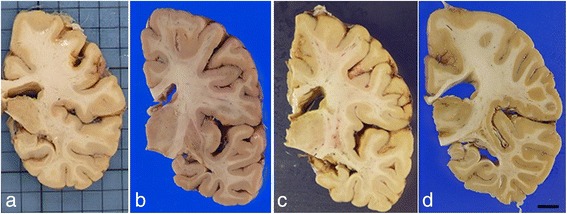
Coronal sections at the basal ganglia and hippocampus level. Cortical ribbons and cerebral white matter are well-preserved. Small cortical infarct in Case 1 (a). a Case 1, (b) Case 2, (c) Case 3, (d) Case 4. Bar = 1 cm
Official websites use .gov
A
.gov website belongs to an official
government organization in the United States.
Secure .gov websites use HTTPS
A lock (
) or https:// means you've safely
connected to the .gov website. Share sensitive
information only on official, secure websites.
Coronal sections at the basal ganglia and hippocampus level. Cortical ribbons and cerebral white matter are well-preserved. Small cortical infarct in Case 1 (a). a Case 1, (b) Case 2, (c) Case 3, (d) Case 4. Bar = 1 cm