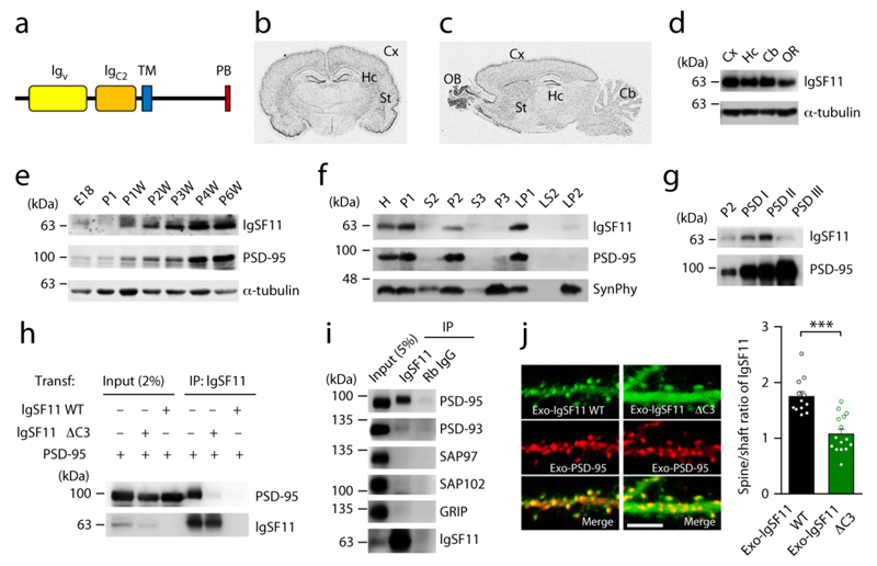
Figure 1. IgSF11 interacts with PSD-95 and is targeted to excitatory synapses in a PDZ interaction-dependent manner.
(a) Domain structure of IgSF11. Igv/C2, immunoglobulin V/C2-like; TM, transmembrane; PB, PDZ-binding.
(b and c) Distribution of IgSF11 mRNA in rat brains (6 weeks) revealed by in situ hybridization. OB, olfactory bulb; Cx, cerebral cortex; St, striatum; Hc, hippocampus; Cb, cerebellar cortex.
(d) Expression of IgSF11 protein in different rat brain regions, by immunoblot analysis. OR, other region.
(e) Expression of IgSF11 protein during rat brain development. E, embryonic; P, postnatal; W, week; PSD-95 and α-tubulin, controls.
(f) Distribution of IgSF11 protein in rat brain fractions. H, homogenates; P1, cells and nuclei-enriched pellet; P2, crude synaptosomes; S2, supernatant after P2 precipitation; S3, cytosol; P3, light membranes; LP1, synaptosomal membranes; LS2, synaptosomal cytosol; LP2, synaptic vesicle-enriched fraction. PSD-95 and synaptophysin (SynPhy; a presynaptic protein) are controls. Three independent experiments were performed.
(g) Detection of IgSF11 in PSD fractions (2 μg of proteins loaded), extracted with Triton X-100 once (PSD I), twice (PSD II), or Triton X-100 and sarcosyl (PSD III). PSD-95 is a control.
(h) IgSF11 forms a complex with PSD-95 in HEK293T cells. IgSF11 ΔC3, a mutant IgSF11 that lacks the last three aa residues and PSD-95 binding; Transf, Transfection; IP, immunoprecipitation. Three independent experiments were performed.
(i) IgSF11 coimmunoprecipitates with PSD-95 in vivo. Deoxycholate (1%) extracts of rat brain crude synaptosomes (6 weeks) were immunoprecipitated and immunoblotted. Gp, ginea pig. Three independent experiments were performed.
(j) IgSF11 is targeted dendritic spines in a PDZ binding-dependent manner. In cultured rat hippocampal neurons transfected with PSD-95 + IgSF11 WT/ΔC3 (DIV14–17), IgSF11 WT shows a greater spine localization (spine/dendritic shaft ratio) than IgSF11 ΔC3. n = 12 (WT) and 15 (ΔC3) neurons, ***p < 0.001, Student’s t-test. Scale bar: 5 μm.

