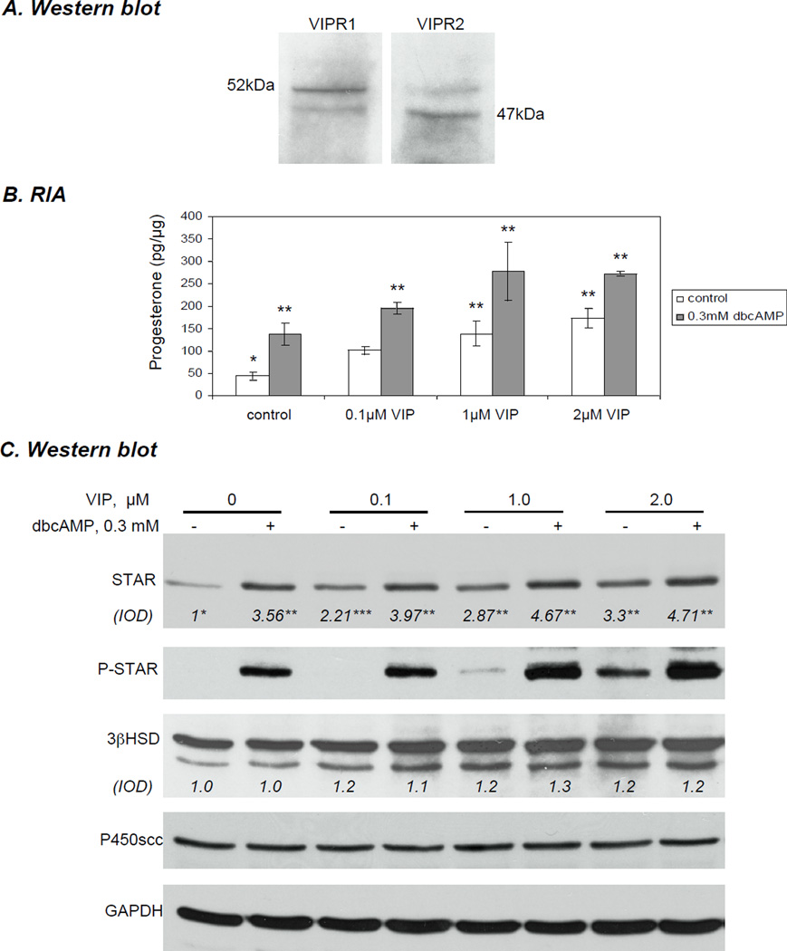
Figure 1.
Expression of VIPR1 and VIPR2 and concentration-dependent increase in STAR expression and steroidogenic output in immortalized KK1 mouse granulosa cells treated with vasoactive intestinal peptide (VIP) against the background PKA activity. Cells were cultured in serum-free DMEM/F12 (1:1) medium with increasing concentrations of VIP with or without 0.3 mM dbcAMP for 6 h. A) Expression of VIPR1 and VIPR2 in KK1 control cells. B) Progesterone production in the collected media was determined by radioimmunoassay C) Cells were collected and homogenized, 20 µg of the lysate was used in western blot analysis of STAR (30 kDa), phospho (P)-STAR (30 kDa), 3β-hydroxysteroid dehydrogenase (3βHSD) (42 kDa), P450scc (45 kDa) and GAPDH (37 kDa). Protein expression was normalised against GAPDH; the average integrated optical density (IOD) for STAR and 3βHSD is shown as fold changes relative to the untreated control.

