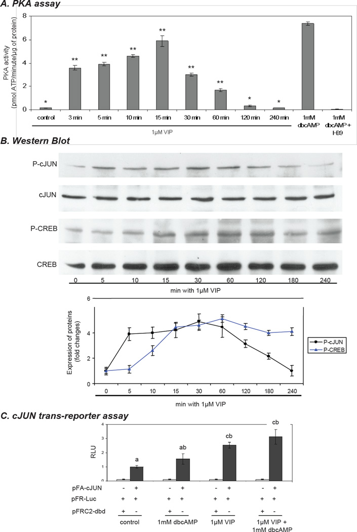
Figure 4.
Time course of vasoactive intestinal peptide (VIP)– induced PKA, CREB and cJUN activation in immortalized KK1 mouse granulosa cells. The KK1 cells were incubated in the presence of 1µM VIP for the times indicated. A) The effect of 1µM VIP on PKA activity in cultured KK1 cells over a period of 4 h. One-way ANOVA with p < 0.0001 and Dunnett’s multiple comparison test were applied; all samples were compared against the control. (**) indicates p < 0.01. B) Representative immunoblots using antibodies against total and phospho (P)- CREB (43 kDa) and (P)- cJUN (48 kDa). The lower panels show the densitometric values (integrated optical density) normalized against the respective total-CREB and -cJUN. One-way ANOVA with p < 0.0001 (CREB; cJUN) followed by Dunnett’s multiple comparison test was applied; all samples were compared against the control indicating: 0’ min. vs 5’-120’ min p<0.01 and 0’ min vs. 180’ min p < 0.05 for P-cJUN and 0’ min vs. 10-240’ min p< 0.01 for P-CREB. C) KK1 cells were transfected with PathDetect cJUN trans-reporting system; 36h after transfection cells were treated with 1 µM VIP and/or 1mM dbcAMP for additional 6 h and luciferase activity in the cell lysate was determined. Lower case letters are used to designate groups that differ significantly (p < 0.01)

