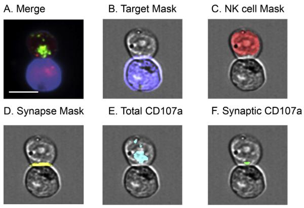
Fig. 3.
Representative masks for analysis of synaptic granules using IFC. Cells were stained as outlined in Section 2 and acquired using IFC. Conjugates were identified using multi-parametric IDEAS algorithm as shown in Fig. 2 and confirmed visually. Masks were developed using IDEAS software to establish regions of interest within each conjugate. A) Example of conjugate identified by IDEAS. Green, CD107a; red, Lysotracker Red; blue, target cell mask. B) Target cell mask (blue): created by using “Morphology” mask that captured all pixels that had fluorescence in target cell. C) NK cell mask (red): created by subtracting target cell mask from total conjugate mask (not pictured). D) Synapse mask (yellow): created by identifying furthest pixels between conjugate mask (not pictured) and target cell mask. E) Total CD107a (cyan): created by using “Intensity” mask to limit region to fluorescent proteins of interest. F) Synaptic CD107a (green): created by identifying pixels common in both the “synapse mask” and “total CD107a” mask. Scale bar = 10 μm. (For interpretation of the references to color in this figure legend, the reader is referred to the web version of this article.)

