Fig. 4.
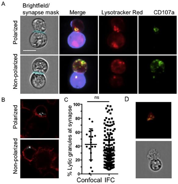
Analysis of conjugates from IFC analysis. Cells were stained as outlined in Section 2 and acquired using IFC. Conjugates were identified using the multi-parametric IDEAS algorithm shown above and confirmed visually with CD107a (degranulated granules, green), Lysotracker Red (lytic granules, red) and Cell Mask Deep Red (target cell stain, purple). Masks were developed using IDEAS software to establish regions of interest within each conjugate. A) Representative examples of polarized (top) and non-polarized (bottom) lytic granules and degranulation B) Representative images acquired by confocal microscopy of NK cells conjugated to K562 targets and fixed, permeabilized and stained for F-actin (phalloidin, red) and perforin as a marker of lytic granules (white). Examples of polarized (top) and non-polarized (bottom) lytic granule localization are shown. C) Percentage of the total NK cell lytic granules localized to the immunological synapse was calculated for IFC and confocal microscopy. p = 0.09 by Student's T-test. Scale bar = 10 μm. D) Representative example of an ex vivo human NK cell conjugated to a K562 target cell. Conjugates were identified using multi-parametric IDEAS algorithm and confirmed visually with CD107a (degranulated granules, green), LysoTracker Red (lytic granules, red). (For interpretation of the references to color in this figure legend, the reader is referred to the web version of this article.)

