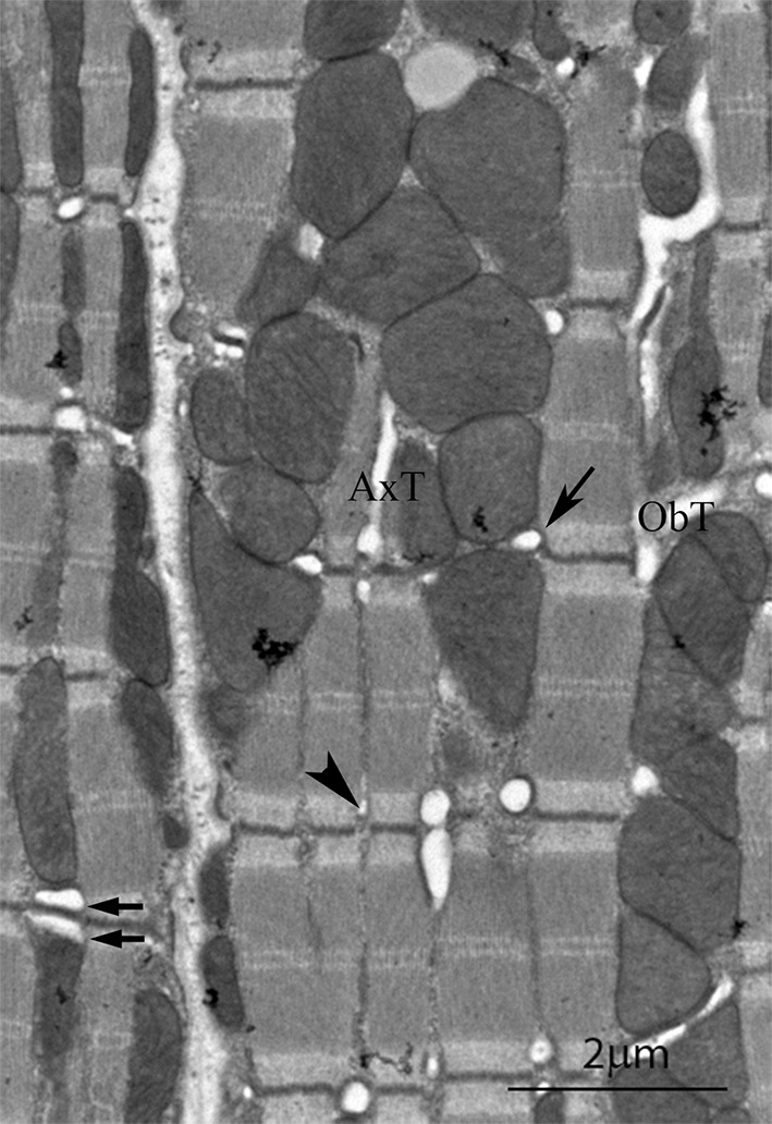
Fig. 1.

Electron micrograph of a longitudinal section of mouse left ventricle showing details of the t tubule system. In the cell on the left the continuity of mitochondrial columns is seen. The cell on the right is sectioned slightly obliquely. (arrow) Transverse tubule of larger diameter at the mitochondria/myofibril boundaries; (arrowhead) smaller diameter tubule at the myofibril/myofibril boundaries. (arrow pair) double transverse tubule; ObT, oblique tubule; AxT, axial tubule
