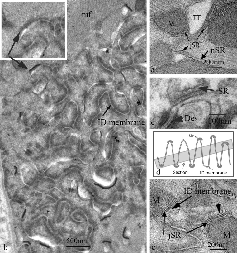
Fig. 4.
a Electron micrograph of mouse papillary muscle showing the organisation of SR, mitochondria (M) and transverse tubules (TT) at a Z-disc. Note the jSR partly sandwiched between the TT and the mitochondria. b Slightly oblique transverse section through a large ID in papillary muscle from a 9 m animal showing the circular profiles of cross sections of the ID folds. No mitochondria are found in this area. The jSR vesicles associated with the ID membrane are shown in red. They are only found at the periphery of the imaged ID. Inset shows indicated area without colour to show the SR vesicle. mf, myofibrils. c High magnification image of jSR vesicle complexed with the top of an ID fold. The SR shows the flat shape and protein-rich regions both within the vesicle and between SR and ID membrane characteristic of cardiac peripheral and t-tubule associated jSR. d Diagram to show that in a slightly oblique section of an ID the SR at the tops of the ID peaks would only be seen at the edges of the imaged ID region. e Electron micrograph showing jSR sandwiched between the ID membrane and mitochondria (M) in longitudinal section. (Color figure online)

