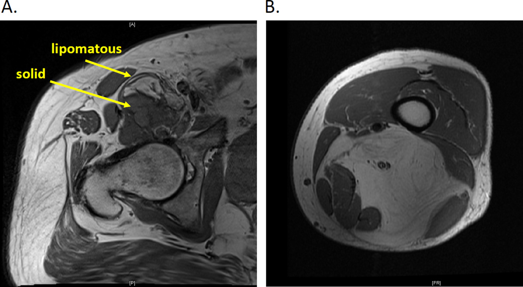
Figure 1.
MRI T1 images showing cross-sectional evaluation of (A) dedifferentiated liposarcoma in the hip and (B) well-differentiated liposarcoma in the thigh. Arrows show lipomatous components representing a well-differentiated component of the dedifferentiated tumor and the nodular high-grade component.

