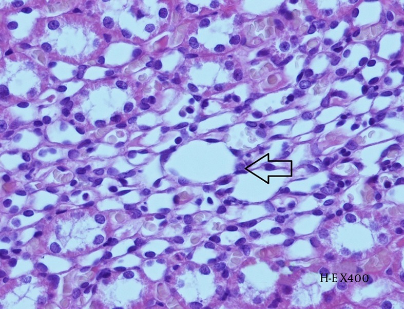
Figure 7. Histopathological Examination of Kidney Tissue From Group N + CIN.

Hydropic degeneration within the tubular epithelium (arrow) and accumulation of contrast agent can be observed in the intertubular space (H-E X 400).

Hydropic degeneration within the tubular epithelium (arrow) and accumulation of contrast agent can be observed in the intertubular space (H-E X 400).