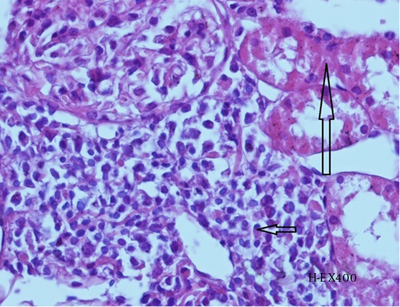
Figure 8. Histopathological Examination of Kidney Tissue From Group CIN.

Mild tubular necrosis (long arrow) and interstitial inflammation (plasma cells- short arrow) can be observed (H-E X 400).

Mild tubular necrosis (long arrow) and interstitial inflammation (plasma cells- short arrow) can be observed (H-E X 400).