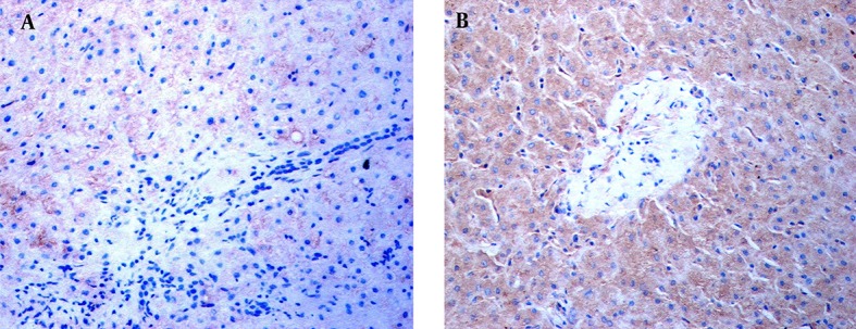
Figure 2. Distribution of Intrahepatic TLR3 Proteins Revealed by Immunochemical Staining.

Immunohistochemical staining was performed using a specific antibody against human the TLR3 protein. TLR3 stains in the livers of an ACH patient and HCC patient are shown. A, No TLR3 stains were found in the NPCs, hepatocytes, or necroinflammatory zone in the liver of the ACH patient at × 400 magnification. B, TLR3 stains with brown granules primarily appeared in the NPCs and hepatocytes, with some staining exhibited in the cells of the portal area, in the HCC subject. The NPCs with TLR3 stains surrounded the hepatocytes at × 400 magnification.
