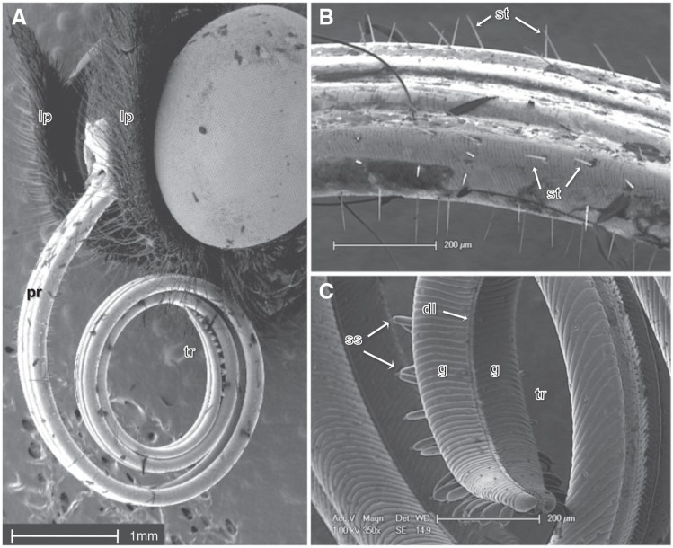
Fig. 1.—.
Scanning electron micrograph of the head and mouthparts of Heliconius melpomene. (A) Labial palps (lp), proboscis (pr) and proboscis tip region (tr). (B) Sensilla trichodea (st) found on the proboscis, (C) Magnified view of the proboscis which is comprised of dorsally and ventrally linked galeae (g) linked by dorsal ligulae (dl). The tip region contains sensilla styloconica (ss) that are club shaped and flattened.

