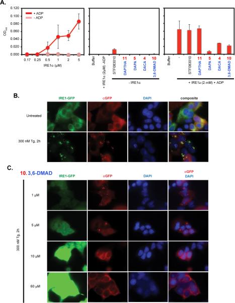
Figure 4. In vitro IRE1α oligomerization and in vivo IRE1α-GFP foci formation assay.
A. Left: Increasing concentration of recombinant IRE1α was used to perform in vitro oligomerization assays with or without 2 mM ADP. Addition of ADP resulted in increased optical density (OD) at higher concentrations of IRE1α, indicating protein oligomerization. Middle: hIRE1α without ADP or each acridine derivative alone do not significantly change optical density readings. STF083010 was used as a control. Right: reactions were performed with hIRE1α plus ADP and 60 μM of inhibitor. B. Immunofluorescent imaging of HEK293 cells overexpressing an IRE1α-GFP fusion protein untreated and treated with 300 nM Tg for 2 hours. IRE1α-GFP foci were detected both through GFP channel and immunofluorescently. DAPI staining was used to visualize nuclei. Anti-GFP/DAPI channels are combined in the composite view on the right. C. The same immunofluorescent imaging as in B with 1-60 μM of Compound 10 for 2 hours.

