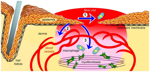
Fig. 1.
Illustration depicts functions of wound epidermis that may be controlled by keratinocyte integrins. Arrow 1 indicates wound re-epithelialization, which is driven by keratinocyte proliferation, local matrix remodeling, and migration. Arrow 2 indicates paracrine signaling from the epidermis to vascular endothelial cells that promotes wound angiogenesis. Arrow 3 indicates paracrine signaling from the epidermis to other wound cells, including inflammatory cells (blue cells) and fibroblasts/myofibroblasts that promote wound contraction (green cells). The wound bed is indicated in red-to-pink gradient.

