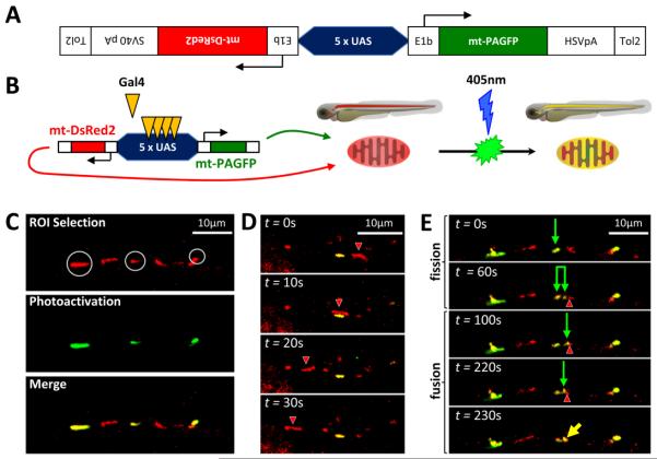
Figure 1. Bidirectional transgene construct enables observation of mitochondrial transport, fission and fusion in dopaminergic neurons in vivo.
A: Schematic depiction of bidirectional transgene construct contained in plasmid pT2-5UAS:mtDsRed2:mtPAGFP
B: Illustration depicting transactivation of construct by Gal4, mitochondrial import and localization of products and photoactivation of mtPAGFP
C, D, E: roy−/−; nacre−/− ; Tg(otpb:gal4); zebrafish embryos were microinjected with pT2-5UAS:mtPAGFP:mtDsRed2 and Tol2 mRNA at the single cell stage. At 3 dpf, single confocal planes were imaged through dopaminergic diencephalospinal axons expressing mtDsRed2 (red) and mtPAGFP (green). (C) Selection and photoactivation of GFP expression in single mitochondria. (D) Timelapse series showing transport of a mitochondrion (red; arrowhead) past a stationary mitochondrion (green). (E) Time-lapse series showing a mitochondrial fission event (green arrow shows parent mitochondrion; paired green arrows show daughter mitochondria), followed by a mitochondrial fusion event (green arrow and red arrowhead show individual mitochondria prior to fusion, yellow arrow shows resulting fusion product).

