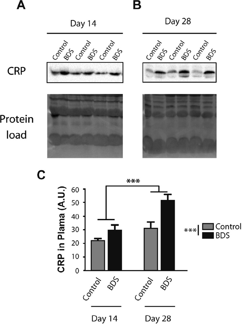
Fig. 4.
Exposure to BDS increases plasma c-reactive protein (CRP) levels in PND14 and PND28 mice. Western blot gels showing levels of CRP in the plasma of PND14 (A) and PND28 (B) mice. Equal protein loading is shown at the bottom (Protein Load) and is the image of activated fluorescent proteins detected by the Stain-Free Gel. (C) Quantification Western blots. Error bars represent mean ± SEM. ***p < 0.0005. N = 7–8 mice in each group.

