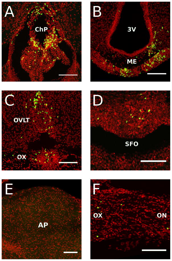
Figure 5.
High resolution digital reconstructed images of Rag2−/− mice reconstituted with 10 million GFP expressing lymphoid cells at 4 weeks after transfer (n = 4 males). A) Choroid plexus (ChP), B) Median eminence (ME), C) Organum vasculosum of the lamina terminalis (OVLT), D) Subfornical organ (SFO), E) Area postrema (AP), F) Optic nerve (on) and chiasm (ox). 3V: Third ventricle. All scale bars = 50 microns

