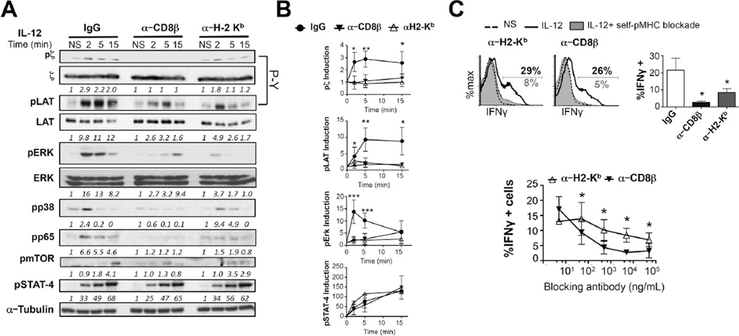
Figure 5. Self-pMHC, and CD8 are required for IL-12-dependent TCR signaling and bystander IFNγ expression.
(A) OT-I CTLs were pre-treated with blocking antibodies against CD8β (5 µg/mL) or H-2Kb (5µg/mL) or isotype controls (IgG) previous to stimulation with 10 ng/mL IL-12. Induction in the levels of pζ (4G10), pZAP-70, pLAT, pERK, p-p38MAPK, p-p65, p-mTOR (S2481) and pSTAT4 were assessed by immunoblot. p21CD3 ζ, LAT and ERK loading was determined following stripping of the same membranes. (B) Densitometry values of each of these phosphoproteins relative to the NS control and normalized to total level of the protein or α-tubulin are shown below the panels or in the graphs. (C) OT-1CTLs from (A) were treated with 5–50,000 ng/mL of the blocking antibodies followed by 6 h of IL-12 stimulation. IFNγ expression was assessed by flow cytometry. All graphs show mean±SD. Data is representative of 3–6 independent experiments. *p < 0.05 **p < 0.005 or ***p < 0.0005. N.S. (non significant).

