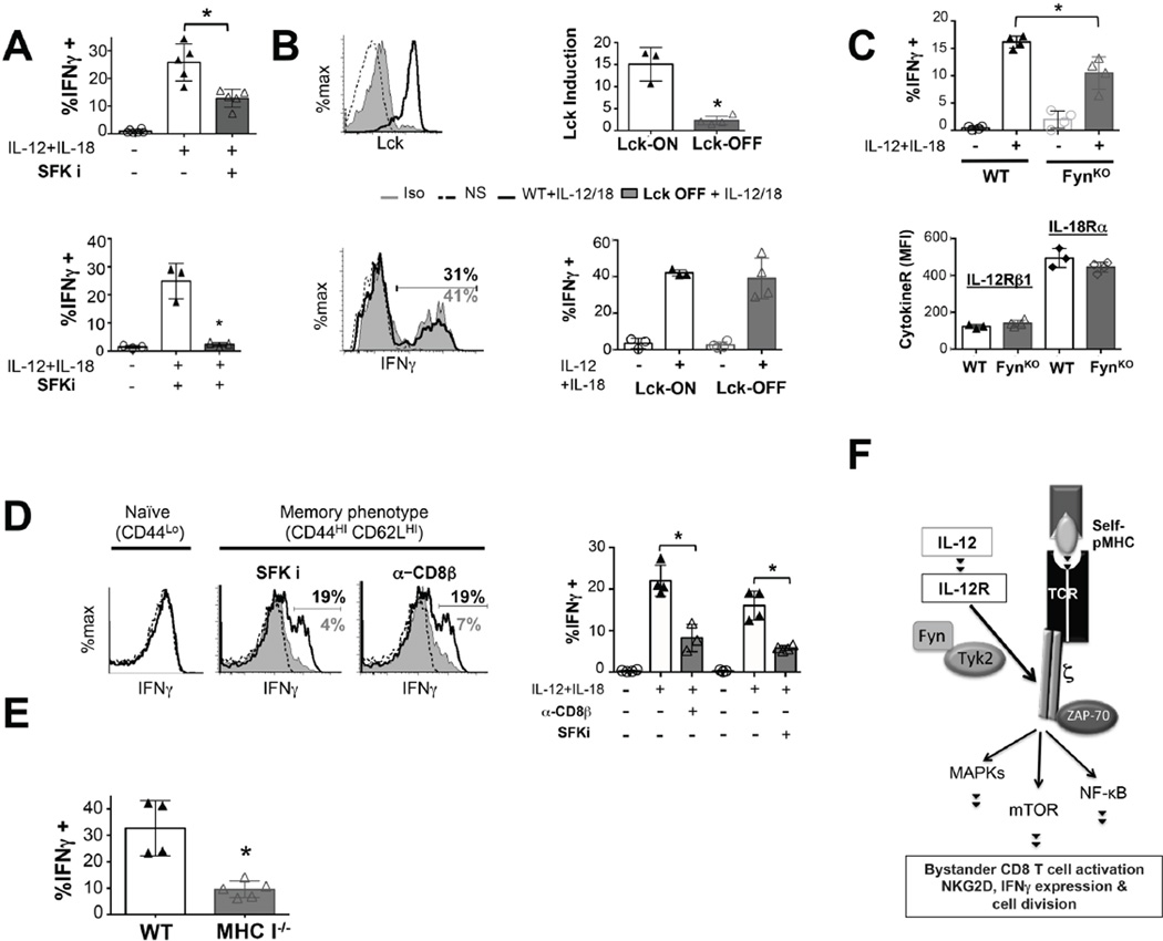
Figure 6. Role of Lck, Fyn and self-pMHC in bystander activation of memory CD8 T cells.
1 × 10⁴ naive OT-I WT, OT-1FynKO or OT-I Lckind naïve cells were adoptively transferred to congenic recipient mice and challenged with LM-OVA (1 × 10⁴ CFU). ≥ 30 days post infection, spleens and lymph nodes were harvested and 1 × 10⁵ OT-1 memory T cells (CD44hi, CD8α+, CD45.2+, Kb-OVA tetramer+) were stimulated for 6h with IL-12+IL-18 (10 ng/mL) and assessed for their IFNγ expression by flow cytometry. (A) OT-1 memory T cells generated in response to LM-OVA (top graph) and (bottom graph) polyclonal CD8 Kb-VSVp (peptide) tetramer positive memory T cells (generated upon challenge of C57BL/6 mice with VSV) were pre-treated with PP2. (B) OT-1Lckind memory cells were treated with doxycycline (Lck-ON) or not (Lck-OFF) overnight in the presence of 5 ng/mL IL-7. Prior to inflammatory cytokine treatment, Lck levels were measured in memory cultures by flow cytometry (top panel). (C) OT-I WT of OT-I FynKO memory cells were assessed for expression of IL-12 and IL-18 receptors and % of IFNγ expressing cells was determined by flow cytometry. (D) CD44hi CD62Lhi memory phenotype CD8 T cells from (32 weeks old) B6 mice were pre-treated with blocking anti-CD8-pMHC (5 µg/mL) or PP2 (10 µM) and stimulated with IL-12 & IL-18 (10ng/mL). 6h later IFNγ was assessed by flow cytometry. (E) 5 × 10⁴ congenically marked polyclonal memory T-cells (CD8-enriched, CD45.1+, CD44hi, Kb-VSVp tetramer+) from ≥ 120 days VSV-infected C57BL/6 mice were adoptively transferred to MHC class I sufficient (WT) or deficient (MHC I −/−) hosts and subsequently re-challenged with (1 × 10⁵ CFU) Listeria monocytogenes. 16 hours later cells were harvested from lymph nodes and spleens and de novo intracellular IFNγ synthesis was measured over 3h. by flow cytometry. All graphs show mean±SD and show MFI values for cytokine receptors or frequency of IFNγ positive bystander activated memory T cells. Data representative of at least 3 independent experiments with n= 3–4 mice per group. * p < 0.05. (F) Model of the mechanism by which IL-12 transduces TCR signals to regulate innate CD8 effector functions. In the presence of self-pMHC-TCR interactions, IL-12 binding to IL-12R allows Tyk2-to activate Fyn, which in turn, phosphorylates CD3ζ to induce TCR-dependent signaling pathways such as ERK, NFκB and mTOR. This enables bystander CD8 functions such as NKG2D or IFNγ expression and cell division.

