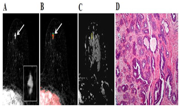
Fig. 3.
49-year-old woman with personal history of right-breast invasive ductal carcinoma, status post lumpectomy. The patient underwent breast MR imaging for high-risk screening. A, Axial dynamic contrast-enhanced initial post-contrast subtraction MR image with magnified inset shows 11-mm lobular homogeneously enhancing mass (arrow) at 11 o’clock of the left breast, 32-mm from the nipple. The lesion has a smooth margin and is at an anterior depth. This lesion was classified as BI-RADS category 4. B, Axial dynamic contrast-enhanced MR image shows the lesion (arrow) has mixed kinetics overall: rapid initial uptake with delayed washout enhancement (red). C, ADC map shows the lesion exhibits low ADC (mean, = 1.37 ×10⁻³ mm2/s), measured within the corresponding region-of-interest (yellow contour). The lesion was classified as fibroadenoma on the basis of D, US-guided core biopsy. Hematoxylin-eosin stain with original magnification, ×100 demonstrates epithelial hyperplasia, high stromal cellularity, no immune cell infiltration and dense stroma.

